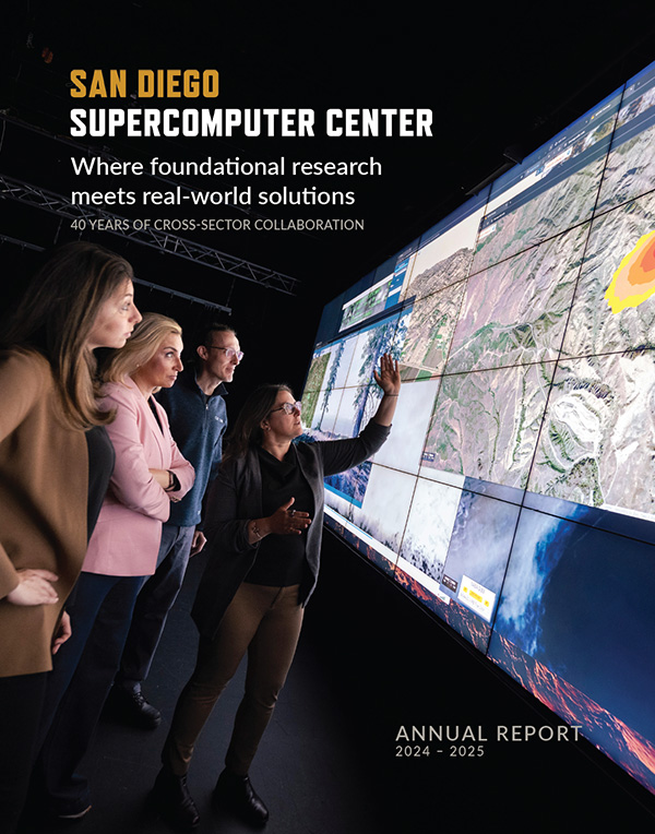
Skip to content

COMPUTATIONAL MEDICINE | Contents | Next

Biochemical Modeling Helps Explain Complex Neural Junction in Chicken Embryo

SERENDIPITOUS COLLABORATION
SYNAPSE SIMULATION
MORE DETAILED GEOMETRY
CONFIRMING A PREDICTION

CSD neurobiologist Darwin K. Berg and his coworkers have achieved a groundbreaking molecular-level description of the functioning of a specialized synaptic junction in the ciliary ganglion of embryonic chicks by combining results from anatomical, neurochemical, microscopic, and computer modeling studies. The study, presented at the annual meeting of the Society for Neuroscience in November 2001, combined the work of Berg’s neurobiology group at UCSD, neuroscientists at UCSD’s National Center for Microscopy and Imaging Research (NCMIR), and computational neurobiologists at the Salk Institute. "We have found this to be a particularly productive collaboration," said Berg. "We knew the power of the microscopy, but we were surprised by the power of the modeling."

somatic spine mat modeled by MCell

Figure 1. Spine Mat Model

This picture of the somatic spine mat modeled by MCell shows neurotransmitter vesicles (large red dots) located at the end of a neuron (not shown). These vesicles release the neurotransmitter acetylcholine, which finds receptors (small blue specks) distributed across the spine mat.

The biochemical modeling program that Berg and his collaborators used is called MCell, which runs on SDSC’s IBM supercomputer, Blue Horizon. The program is also at the center of an NPACI alpha project and an NSF Information Technology Research project led by NPACI Director Fran Berman. "I think we’ve shown that MCell is becoming a valuable means of checking on the results of chemical, microscopic, and electrophysiological studies of complex events occurring rapidly on the scale of microns to millimicrons," said Salk researcher Thomas Bartol. The ciliary ganglion is a small tangle of neurons that supplies nerve connections to the muscle controlling the pupil of the eye and to muscle in the eyeball. Berg, graduate student Richard Shoop, and postdoctoral researcher Jay Coggan spent several years characterizing the fine structure and anatomy of their target junction, elucidating the detailed functioning of neurotransmission across it. "We were studying the ciliary ganglion of the chick embryo because vision systems in birds are both well developed evolutionarily and more straightforward to study than in mammals," said Berg. "The ciliary ganglion is an exemplary form of a complex type of neural junction, quite distinct from the fine, dendritic junctions of brain neurons and from the much larger and coarser neuromuscular junctions found in other parts of the body.

SERENDIPITOUS COLLABORATION

"We had been studying it for several years as an interesting class of hybrid vertebrate synapses," Berg said. His group was using its own well-equipped chemical lab, while also taking advantage of electron microscopes available in the UCSD Division of Neurobiology. "At one point, a few years ago, those microscopes went down for a rebuild," Berg said. "That was when we turned to NCMIR," in the lab of UCSD Medical School neuroscientist Mark H. Ellisman. "We became aware of MCell almost by accident," Berg said.

Figure 2. A Postsynaptic Density

This pair of images highlights activity in the region of the postsynaptic density (black circular area) 10 microseconds (upper image) and 200 microseconds (lower image) after release of acetylcholine (small green ovals) from a synaptic vesicle (large red dot). Alpha-3 receptors (triangles) predominate in the postsynaptic density area while alpha-7 receptors (pentagons) are distributed outside this area. Colors indicate state of activation–prereceptive (blue), activatible (red, green, and brown), fully activated (yellow and orange), or deactivated (black). Diameter of the postsynaptic density area is about 0.5 microns.

"We were delighted to work with the Berg group," said NCMIR Associate Director Maryann E. Martone. The NICMIR researchers had just finished a study of the large-scale neuromuscular junctions in the body–also modeled by Bartol and the group at Salk–and Berg’s smaller and more complex target seemed a logical next step. NCMIR, funded by the National Institutes of Health’s National Center for Research Resources, introduced Berg’s group to their 400 keV electron microscope, which allows study of much thicker sections of tissue than those Berg had been using across the campus. "These make 3-D reconstruction of the tissue sections much easier," said Martone. NCMIR scientists have worked with a constellation of other scientists, including many from SDSC, to link the powerful electron microscope with a computational system in their own lab (now part of the Keck I satellite cluster of SDSC). NCMIR has also pioneered the development of programs for visualizing neuronal structure in three dimensions, using accurate computational reconstructions of whole neurons from serial sections.

A complete 3-D reconstruction of a portion of an embryonic chick ciliary ganglion was carried out by the collaboration in 1999 and 2000. The reconstruction showed the somatic spines, branching structures for receiving neural signals that emerge from and are tightly folded against the soma (body) of the neuron (Figure 1). "Somatic spines grouped in discrete clumps or mats are the hallmark of this kind of junction," Berg said. In the reconstruction, arbitrary colors were used to distinguish one spine from another, "but they almost certainly function together as a group," he said.

SYNAPSE SIMULATION

Figure 3. Close-Up of Spine Mat

The location of two overlying vesicles is represented by the red dots. In this close-up, the neurotransmitter acetylcholine (small green ovals) was released from the upper vesicle 200 microseconds ago. The alpha-7 receptors (pentagons) are more numerous in areas outside of postsynaptic densities, while alpha-3 receptors (triangles) are scarce here (see Figure 2 color code). The distance across the image is 0.5 microns.

Neuromuscular junctions of all types are able to deliver high-
frequency, reliable stimuli to the postsynaptic muscle fiber. In the chick ciliary ganglion, the neurotransmitter acetylcholine is released from a calyx, so called because the tiny structure is reminiscent of a flower part having the same name. The calyx envelops and surrounds the somatic bodies of the ganglion and overlies the mats of spines.

In their chemical studies, Berg and his group had found that the spine mat surfaces have two types of receptors for the neurotransmitter acetylcholine. One type, carrying a sequence called the alpha-7 gene product, is particularly permeable to calcium ions, allowing them into the cell, where they regulate many processes. The other, called alpha-3, is much less permeable to calcium. Paradoxically, the alpha-7 receptors were widely distributed across the somatic spines, while the alpha-3 receptors occupied only a few small areas called postsynaptic densities–key entryways into the cell. "We knew that both types of receptor were involved in neurotransmission across the ganglion," Berg said, "but we needed to know exactly how they functioned–together or in competition with one another." Martone and Ellisman suggested that the Berg group consider using Bartol’s MCell code to model the detailed, dynamic, time-dependent behavior of the receptors.

MORE DETAILED GEOMETRY

Bartol also was excited by the prospect. "MCell does detailed biophysical modeling, and the ciliary ganglion seemed an ideal next project to work on after the larger neuromuscular junction project," he said. The researchers went from the visually reconstructed spine mat covered with receptors to a much more detailed geometry. "We had to put numbers on it," Bartol said. This massive task was undertaken by Eduardo Esquenazi, a recent UCSD graduate working in Ellisman’s lab. Under the direction of Coggan, Martone, and NCMIR staff scientist Naoko Yamada, Esquenazi performed detailed measurements of the electron microscope data and added the chemical information from Berg’s lab.

Computationally, the geometric reconstruction for MCell also took advantage of the capabilities of a code called Xvoxtrace, developed by Stephan Lamont, a computational visualization specialist in Ellisman’s laboratory. Lamont worked with Bartol, Esquenazi, and others on the project to make the necessary modifications to Xvoxtrace.

CONFIRMING A PREDICTION

Some of the results of the modeling with MCell are shown in Figures 2 and 3. "What we found helped to confirm our idea that the two receptor types operate both cooperatively and competitively to modulate the overall response to neural signal in a way that makes the entire ciliary ganglion extremely sensitive," said Berg. Thus, it is able to modulate the opening and closing of the chick pupil in response to changing light levels. "Despite the fact that the junction appears as a rather messy set of spine mats, its overall functioning can be rapid and sensitive enough to permit very fine control of the pupil," Berg said.

The MCell model allows "parameter sweep" studies evaluating the neurochemical behavior of complex systems. "In the dynamic modeling," Bartol said, "we can estimate many parameters that would otherwise be very difficult to determine, like the rate of transmitter-receptor binding at each point." Martone noted that the study points the way to a new kind of multi-laboratory collaboration that can synthesize the results of very different kinds of study–anatomical, neurochemical, microscopic, and modeling. "It was a perfect problem for NCMIR, because it pushed us to develop new tools," she said.

"MCell gives us another way to obtain biological answers to fundamental questions," said Berg. "We had predicted that the receptors would work in certain ways, and the modeling provided a spectacular confirmation." –MM


Project Leader 
Darwin K. Berg, UCSD

Participants
Jay S. Coggan,
Mark H. Ellisman,
Eduardo Esquenazi,
Stephan P. Lamont,
Maryann E. Martone,
Richard D. Shoop,
Naoko Yamada
UCSD

Thomas M. Bartol,

Terrence J. Sejnowski
Salk Institute
s
References

Shoop, R. D., E. Esquenazi, N. Yamada, M. H. Ellisman, and D. K. Berg (2002): Ultrastructure of a somatic spine mat for nicotinic signaling in neurons, Journal of Neuroscience 22: 748-756

E. Esquenazi, J. S. Coggan, T. M. Bartol, R. D. Shoop, T. J. Sejnowski, M. H. Ellisman, D. K. Berg (2001): Computer simulation of synaptic ultrastructure and microphysiology in the chick ciliary ganglion, Society for Neuroscience annual meeting poster.